Kansas State University

search

K-State Turf and Landscape Blog

Tag: diagnostic lab

Got Plant Questions? The K-State Plant Disease Diagnostic Lab Has Answers.

By Chandler Day, Associate Diagnostician, K-State Plant Disease Diagnostic Lab


A quality diagnoses starts with a quality sample. A sample can be physical plants or digital images of plants. Digital images are a great screening tool to determine if a physical sample is required for diagnoses. In order to make a diagnosis, the quality of the images and/or samples is extremely important. Follow these tips for submitting your plant health questions. If you ever have questions about a plant problem, how to collect and/or ship a sample, feel
free to call or email us.

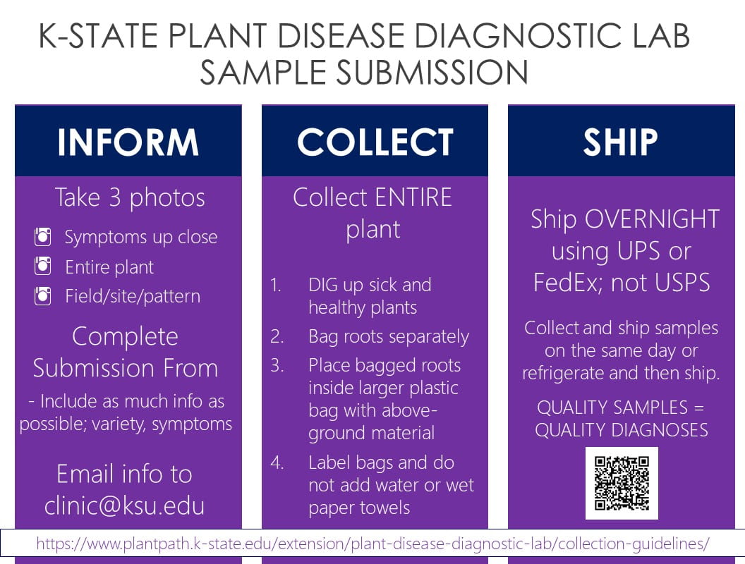
Inform. Collect. Ship.

Inform:

Send photos and background information to clinic@ksu.edu!

Three types of high quality digital images are an excellent way to pre-screen plant health issues.

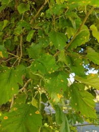
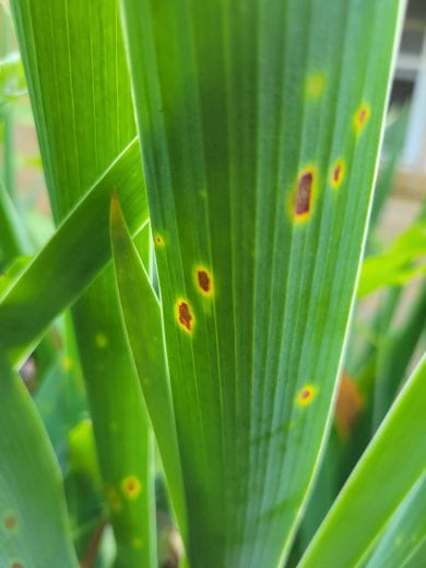
  1. Symptoms up close (Fig. 1)
  • Take zoomed in photos of the problem/symptoms.
  • Ensure the image is in focus by tapping the image before you take the shot.
  • Examples of symptoms: leaf spot, branch die-back, sunken tissue, scorched leaves.
Fig 1. Close-up and focused image of leaf spots on iris.


  1. Entire plant. (Fig. 2.)
  • Take photos of the entire plant that includes all plant parts from soil level to the top of the plant.
  • Ensure the symptoms are still visible in this type of image.
  • Example: whole tree or shrub (trunk/base to top of crown).


Fig. 2. Photo of entire Iris plant with leaf spot still visible.


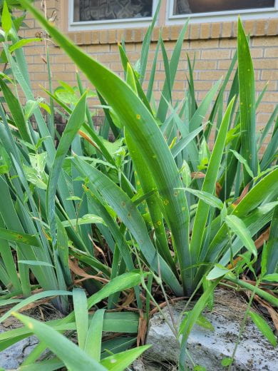
  1. Landscape pattern. (Fig. 3.)
  • Stand back and capture the entire landscape where the plant resides.
  • Include in the photo the surrounding plants, concrete, rocks, drain spouts, or whatever else is near the symptomatic plant. Don’t worry if the symptoms cannot be seen in this type of image.
  • The importance of this image is not to capture the symptoms on the plant but to capture the landscape. This gives us look at how the affected plant is growing within the site and if there are any site issues that might be contributing to the problem.
Fig. 3. Overall landscape view of flowerbed with Iris plants.


Useful background information:

  1. Site history:
    • Soil types, drainage, slope, sunny or shady problem areas, previous construction activity, proximity to structures such as roads or sidewalks, etc.
  2. Irrigation practices:
    • Frequency of irrigation, length of time, irrigation application method (sprinkler, drip, hand held hose), time of day
  3. Chemical history:
    • Pesticide usage and timing, fertilizer applications, etc.
  4. Pattern on plant:
    • Describe the problem. Are symptoms on new or old growth? Top or bottom of plant?
  5. Pattern in landscape:
    • One host or multiple hosts? Other plants in the landscape showing similar symptoms?
  6. Timing:
    • When did the symptoms occur: All at once? (i.e. after a storm?) Slowly over time?


Collect:

  • Complete the sample submission form with as much information as possible.
  • Send a “healthy” plant and a “sick” plant.
  • Submit entire plants when possible including roots. (EX. Tomatoes, annuals, turf grass, etc.)
  • DIG up plants. Do NOT pull up plants as this can damage the roots.
  • Bag roots separately and then place entire plant into larger plastic bag.
  • Do NOT add water or use paper bags. These degrade the sample quality and affects the diagnostic process.
  • For specific collection guidelines, go to the K-State Plant Disease Diagnostic Lab webpage, scroll down to where it says collection tips, and use the arrows on the right hand side to navigate to the appropriate collection strategy.


Ship:

  • Collect and ship samples on the same day. If this is not possible, store plants in plastic bags in the refrigerator until shipping is possible.
  • Ship plants overnight using UPS or FedEx. UPS can take up to 14 days even with 2 day priority shipping.
  • Ship on or before Wednesday to avoid weekend storage.


The K-State Plant Disease Diagnostic Lab is here to help you identify your plant health problems. If you ever have questions about a plant problem, how to collect and/or ship a sample, feel free to call or email us.


K-State Plant Disease Diagnostic Lab
4032 Throckmorton PSC
1712 Claflin Road
Manhattan, KS 66506
clinic@ksu.edu
785-532-6176


Rose Rosette Virus Active in Summer 2023

By Judy O’Mara, Director, K-State Plant Disease Diagnostic Lab


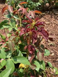
Rose rosette is currently showing up in Kansas gardens. This disease is a serious problem in wild multiflora roses but also goes to many common garden roses.

The rose rosette virus is spread from plant to plant by a microscopic eriophyid mite (Phyllocoptes fructiphilus).  It is the only known insect vector of the rose rosette disease. The mite can be wind dispersed or moved around on tools, clothing or infected plant material. Mites can successfully overwinter on the rose plant. The rose rosette virus can also be spread through grafting.

Rose rosette causes a progressive deformity of the rose plant that gets worse over time. It is challenging to identify visually because symptoms vary widely from plant to plant. Some prominent symptoms can include reddish or distorted leaves, stems that are elongated and thickened, a proliferation of shoots (rosette), and excessive thorniness. Plants infected with rose rosette are also more susceptible to winter damage.

An initial symptom of rose rosette might be an elongated stem with reddish 
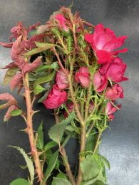
leaves that stands out above the normal growth habit of the shrub. Reddish leaves are a tricky symptom because roses put out new flushes of growth throughout the growing season and the new leaves commonly start out red and then green up.

More characteristic for rose rosette virus are symptoms of bunchy growth or a proliferation of shoots. This symptom is referred to as a rosette or witches’ broom.  Some roses have a bunchy growth pattern so keep in mind that this symptom development should look new or different from the established growth habit of the plant.

A striking symptom for rose rosette is excessive thorniness along the stems. These thorns are generally small, green and soft whereas normal thorns tend to be larger, hard and sharp. Infected plants usually have both types of thorns present.

Pruning out symptomatic portions of infected rose plants will not eliminate the disease.

Plants with rose rosette can survive for one to two years but the virus will continue to spread within a planting. It is important to remove infected plants as soon as possible, including the roots. Infected  plants should be placed in trash bags to reduce further spread of the eriophyid mites.

Interspersing roses with non-rose hosts can slow down the spread of the mite and the rose rosette disease. Dead heading roses throughout the growing season will help minimize the habitat of the eriophyid mite. Resistance is not available for commonly grown garden roses. Native roses resistant to rose rosette include: prickly rose (Rosa acicularis ), prairie rose (Rosa arkansana), smooth rose (Rosa blanda), swamp rose (Rosa paulustris), Carolina rose (Rosa carolina), and scotch rose (Rosa spinosissima).

Visual identification of rose rosette virus disease can be challenging. The K-State Plant Disease Diagnostic Lab does offer a PCR test that can confirm the presence of rose rosette virus.  Testing can be expensive but might be worthwhile in locations with large plantings of roses.


K-State Plant Disease Diagnostic Lab

4032 Throckmorton PSC
1712 Claflin Road
Manhattan, KS 66506
clinic@ksu.edu
785-532-6176


Useful references:

Rose Rosette Disease: A Diagnostic Guide. N. Claros, etal. https://apsjournals.apsnet.org/doi/10.1094/PHP-05-22-0047-DG

Rose Rosette Disease. J. Olson, etal.
https://extension.okstate.edu/fact-sheets/rose-rosette-disease.html#management-guidelines-for-rrd

Tips for submitting a ‘digital sample’ to plant disease diagnostic lab

By Lucky Mehra

When it comes to plant health, physical samples are best. However, sometimes it is not practical to send physical samples, such as with large trees or shrubs. A digital sample can be a good alternative or a good first step. By ‘digital sample’ we mean submitting digital images of the plant problem to the KSU plant disease diagnostic clinic.

Consider the following tips to take the photos and provide all the relevant information to help us diagnose the problem quickly and correctly.

Pictures

The main component of a ‘digital sample’ is the set of digital images itself. Take the following types of pictures to help us understand both the ‘big’ and ‘small’ picture of the problem. Make sure that the plant or plant part is in focus when taking pictures. Some phones are pretty good at auto focus, but most of the time you will need to tap on the screen at the point of interest to guide your camera to focus at a particular point.

  • Take pictures from a distance to give us the landscape view or the ‘big picture view’. It should include the whole tree/shrub along with neighboring plants (Fig. 1).

Fig. 1. The Hawthorn tree in the foreground has leaf spots. This image is to give an overall view of the landscape. For example, proximity of the tree to the concrete and adjoining trees.

  • Photo of the affected plant(s) i.e. the plant(s) showing the problem.
  • Photo of the affected plant part, whether it is leaf, flower, stem, twig, or root (FIg. 2).

Fig. 2. Affected leaves of the hawthorn tree.

  • Try to take a close-up shot of the problem symptoms (Fig. 3). If there are signs (actual parts of the pathogen i.e. fungal mycelium or other structures) present on the plant part, try to zoom in, tap to focus, and then take the photo. Take multiple photos to ensure that you will have at least a few good quality ones. If you have access to a microscope, you can bring the affected plant part to your home or office and focus it under the microscope. With some patience, you can also take really good macro shots with your phone through the eyepiece lens. Many of our students take photos this way in the lab. 

Fig. 3. An example of close-up images of a hawthorn leaf (top side of leaf on the left, and underside of leaf on the right) with yellow spots taken with a phone camera, without any additional lens attachment.

Additional essential information

Sometimes, the plant problem is very peculiar, and it is easy to identify by just looking at the photos you submit (e.g. yellow spots on hawthorn leaves shown above are the symptoms of Cedar-hawthorn rust); however, most of the time it is not possible to diagnose a problem only based on photos. We need much more information from you to help us with the correct diagnosis. Provide as much information as you can. Please see below for the type of information that will be useful to us when making the diagnosis.

Site history and pattern

Make sure to provide information about the site where the shrub or tree is located. That information may include soil type if known, soil pH, slope, distance to the concrete sidewalk or road etc. 

Are there any drainage problems? Is this the only plant affected? Are other plants of the same species affected? If yes, what is the pattern of these plants in the landscape i.e. is there a cluster of affected plants or the distribution is random? Are other plant species affected?

Plant pattern

Tell us about the affected plant part whether it is leaf, stem, flower, twig, stem or root. Additionally, report the location of symptoms on the plant. Some problems tend to occur on younger leaves, others are specific to older leaves. All this information can help us rule out some issues and narrow down the diagnosis.

Timing of the symptom appearance.

Was there any weather event such as temperature (too hot or too cold) or moisture extremes (e.g. heavy rainfall) prior to the symptom development. We can download these data from a local weather station as well, however, rainfall events can be non-uniform over the whole area covered by a weather station. So if you have onsite information about the weather data, report it to us.

Chemical history

Any history of chemical or fertilizer application should be provided. This can help the diagnostician in figuring out if the problem is arising due to chemical exposure or due to a biotic agent.