Kansas State University

search

K-State Turf and Landscape Blog

Category: Diseases

Tips for submitting a ‘digital sample’ to plant disease diagnostic lab

By Lucky Mehra

When it comes to plant health, physical samples are best. However, sometimes it is not practical to send physical samples, such as with large trees or shrubs. A digital sample can be a good alternative or a good first step. By ‘digital sample’ we mean submitting digital images of the plant problem to the KSU plant disease diagnostic clinic.

Consider the following tips to take the photos and provide all the relevant information to help us diagnose the problem quickly and correctly.

Pictures

The main component of a ‘digital sample’ is the set of digital images itself. Take the following types of pictures to help us understand both the ‘big’ and ‘small’ picture of the problem. Make sure that the plant or plant part is in focus when taking pictures. Some phones are pretty good at auto focus, but most of the time you will need to tap on the screen at the point of interest to guide your camera to focus at a particular point.

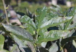
  • Take pictures from a distance to give us the landscape view or the ‘big picture view’. It should include the whole tree/shrub along with neighboring plants (Fig. 1).

Fig. 1. The Hawthorn tree in the foreground has leaf spots. This image is to give an overall view of the landscape. For example, proximity of the tree to the concrete and adjoining trees.

  • Photo of the affected plant(s) i.e. the plant(s) showing the problem.
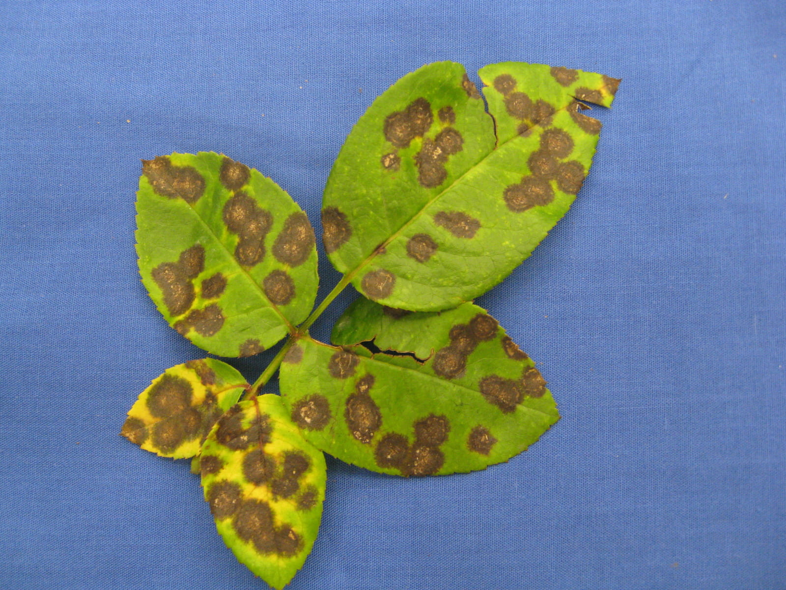
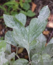
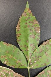
  • Photo of the affected plant part, whether it is leaf, flower, stem, twig, or root (FIg. 2).

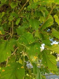
Fig. 2. Affected leaves of the hawthorn tree.

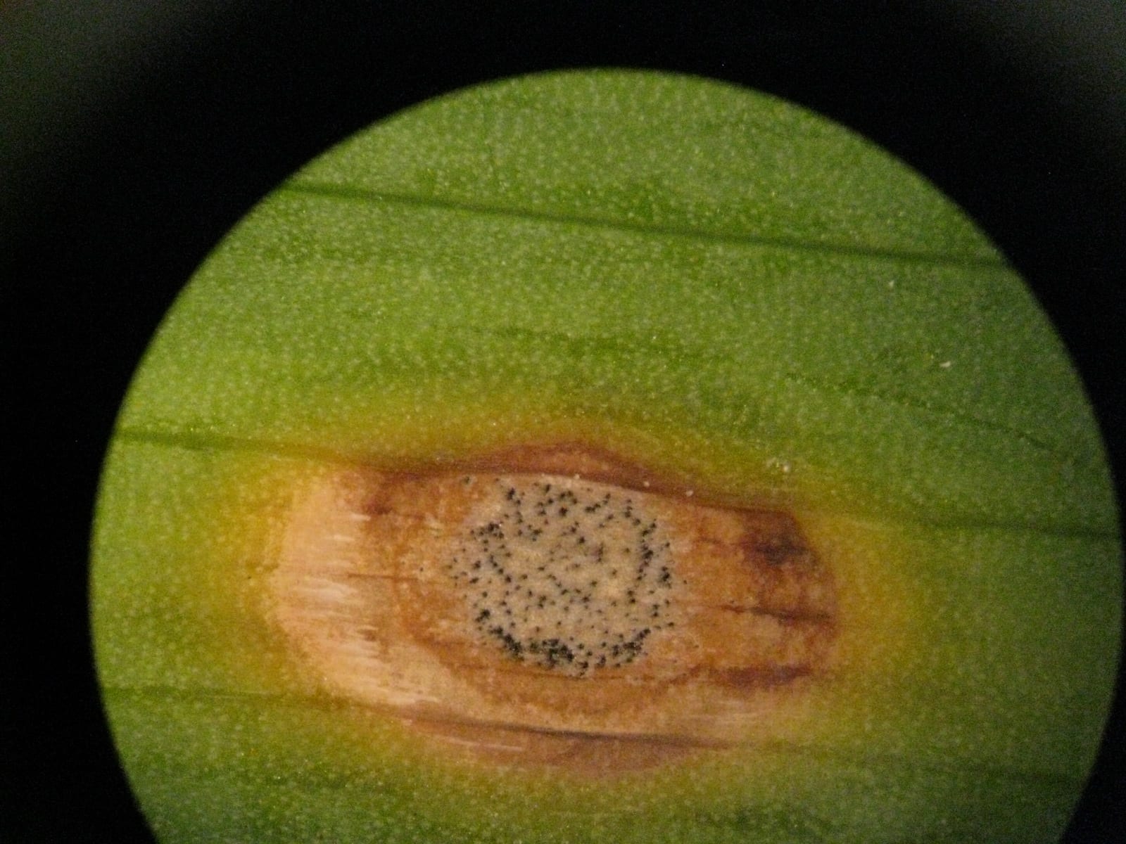
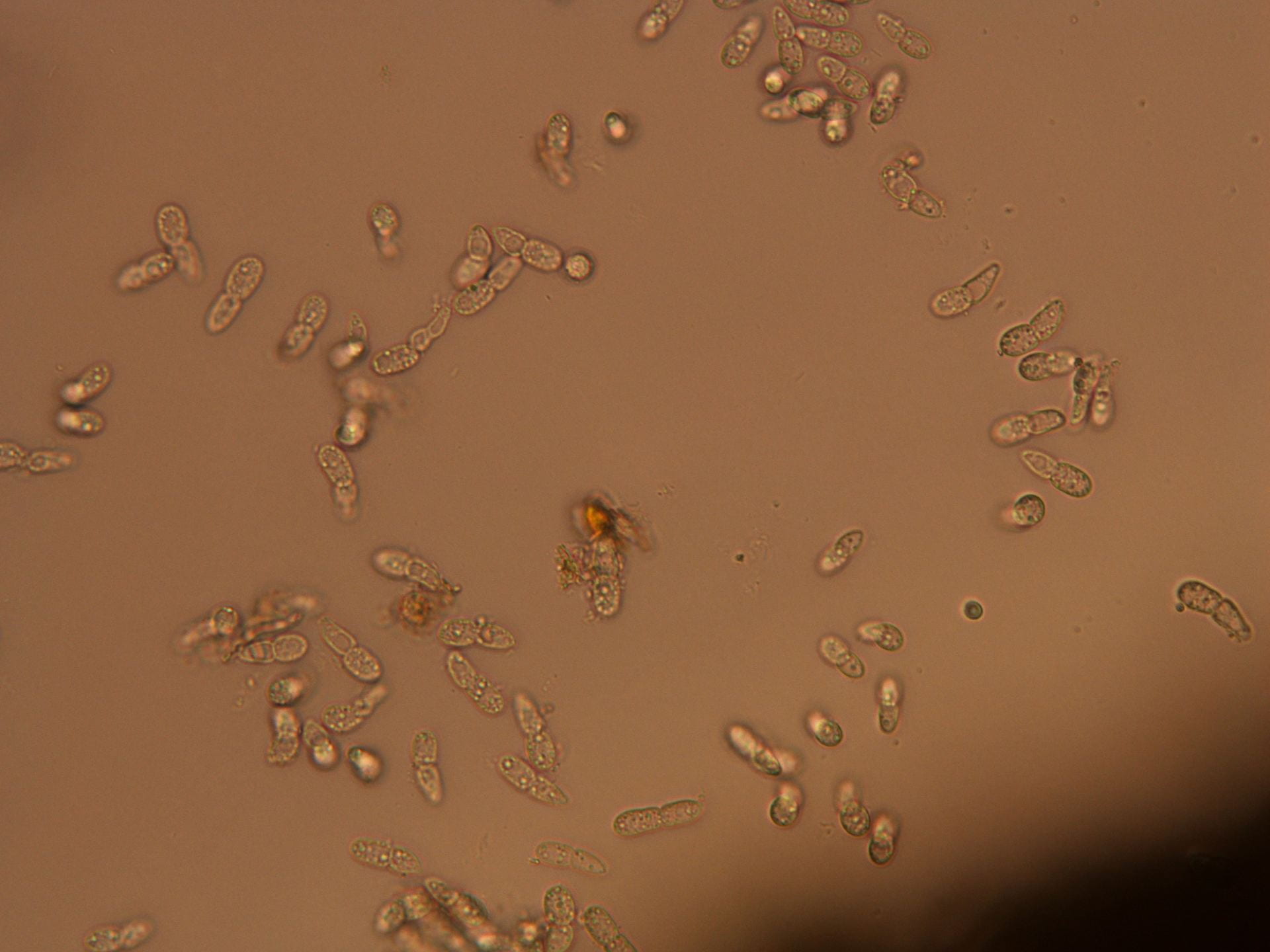
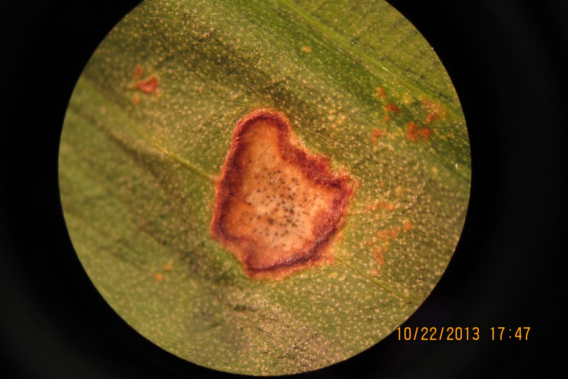
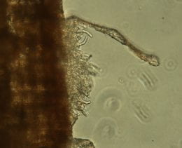
  • Try to take a close-up shot of the problem symptoms (Fig. 3). If there are signs (actual parts of the pathogen i.e. fungal mycelium or other structures) present on the plant part, try to zoom in, tap to focus, and then take the photo. Take multiple photos to ensure that you will have at least a few good quality ones. If you have access to a microscope, you can bring the affected plant part to your home or office and focus it under the microscope. With some patience, you can also take really good macro shots with your phone through the eyepiece lens. Many of our students take photos this way in the lab. 

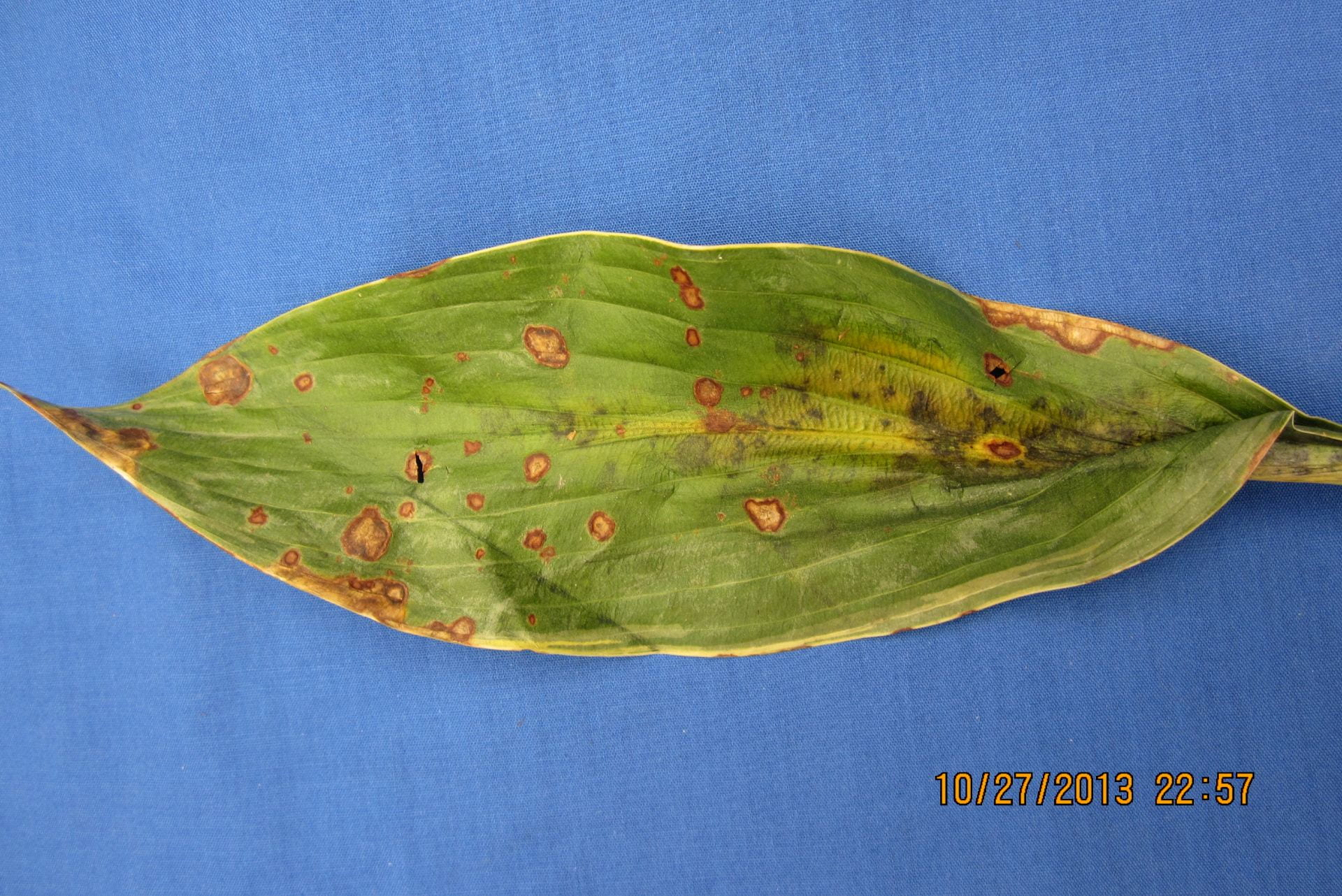
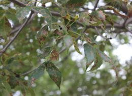
Fig. 3. An example of close-up images of a hawthorn leaf (top side of leaf on the left, and underside of leaf on the right) with yellow spots taken with a phone camera, without any additional lens attachment.

Additional essential information

Sometimes, the plant problem is very peculiar, and it is easy to identify by just looking at the photos you submit (e.g. yellow spots on hawthorn leaves shown above are the symptoms of Cedar-hawthorn rust); however, most of the time it is not possible to diagnose a problem only based on photos. We need much more information from you to help us with the correct diagnosis. Provide as much information as you can. Please see below for the type of information that will be useful to us when making the diagnosis.

Site history and pattern

Make sure to provide information about the site where the shrub or tree is located. That information may include soil type if known, soil pH, slope, distance to the concrete sidewalk or road etc. 

Are there any drainage problems? Is this the only plant affected? Are other plants of the same species affected? If yes, what is the pattern of these plants in the landscape i.e. is there a cluster of affected plants or the distribution is random? Are other plant species affected?

Plant pattern

Tell us about the affected plant part whether it is leaf, stem, flower, twig, stem or root. Additionally, report the location of symptoms on the plant. Some problems tend to occur on younger leaves, others are specific to older leaves. All this information can help us rule out some issues and narrow down the diagnosis.

Timing of the symptom appearance.

Was there any weather event such as temperature (too hot or too cold) or moisture extremes (e.g. heavy rainfall) prior to the symptom development. We can download these data from a local weather station as well, however, rainfall events can be non-uniform over the whole area covered by a weather station. So if you have onsite information about the weather data, report it to us.

Chemical history

Any history of chemical or fertilizer application should be provided. This can help the diagnostician in figuring out if the problem is arising due to chemical exposure or due to a biotic agent.

Iris Leaf Spot: A Common Problem in Kansas

By Judy O’Mara

I was out working in the garden last weekend and I saw the first appearance of iris leaf spot (Didymellina macrospora). This is a very unattractive fungal disease. The iris planting puts out nice looking flowers each year but for the rest of the season everything looks rough, with heavy leaf spotting and leaf scorch. Iris leaf spot will show up in most years but will be severe in years that are wet.

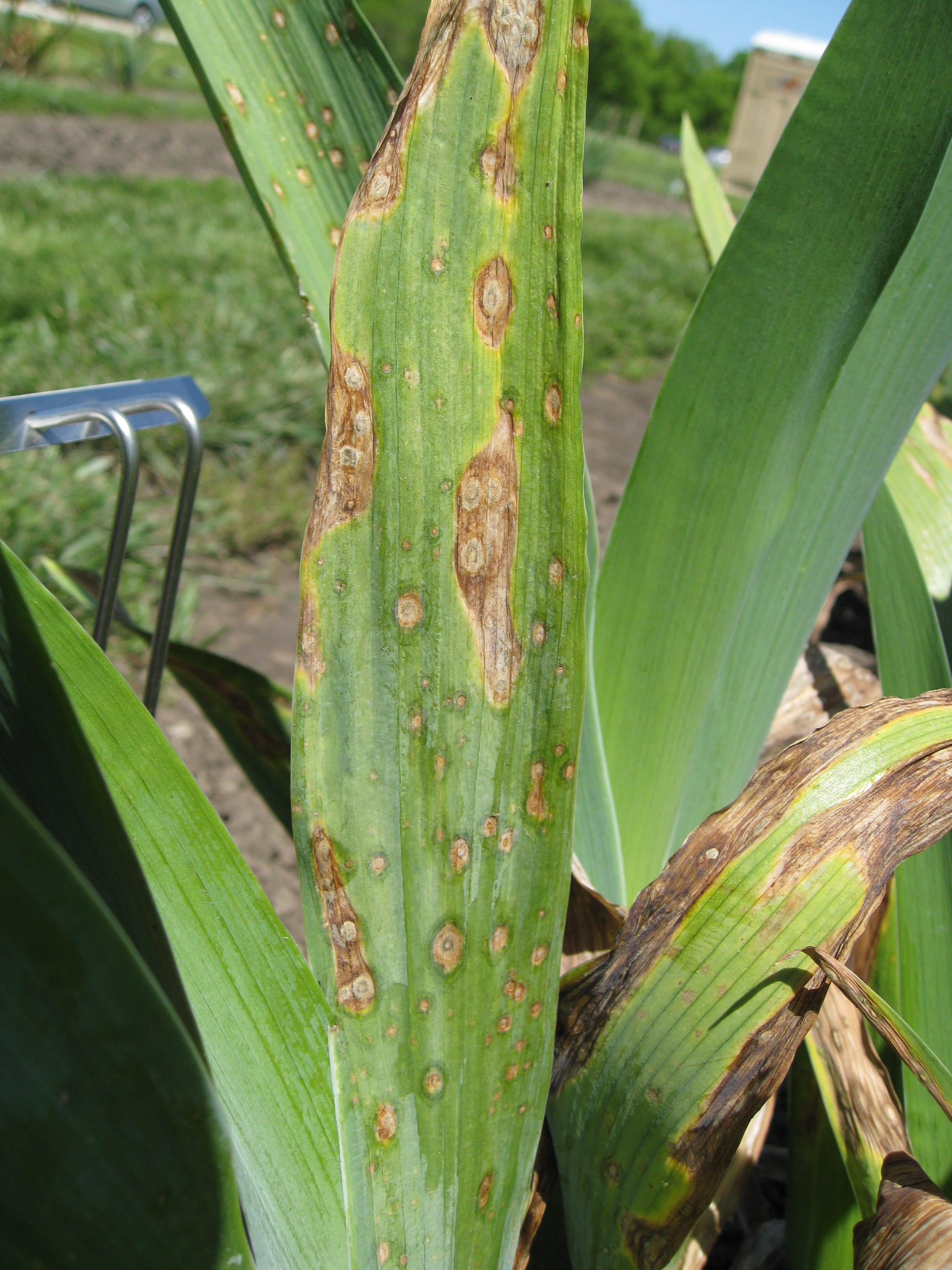
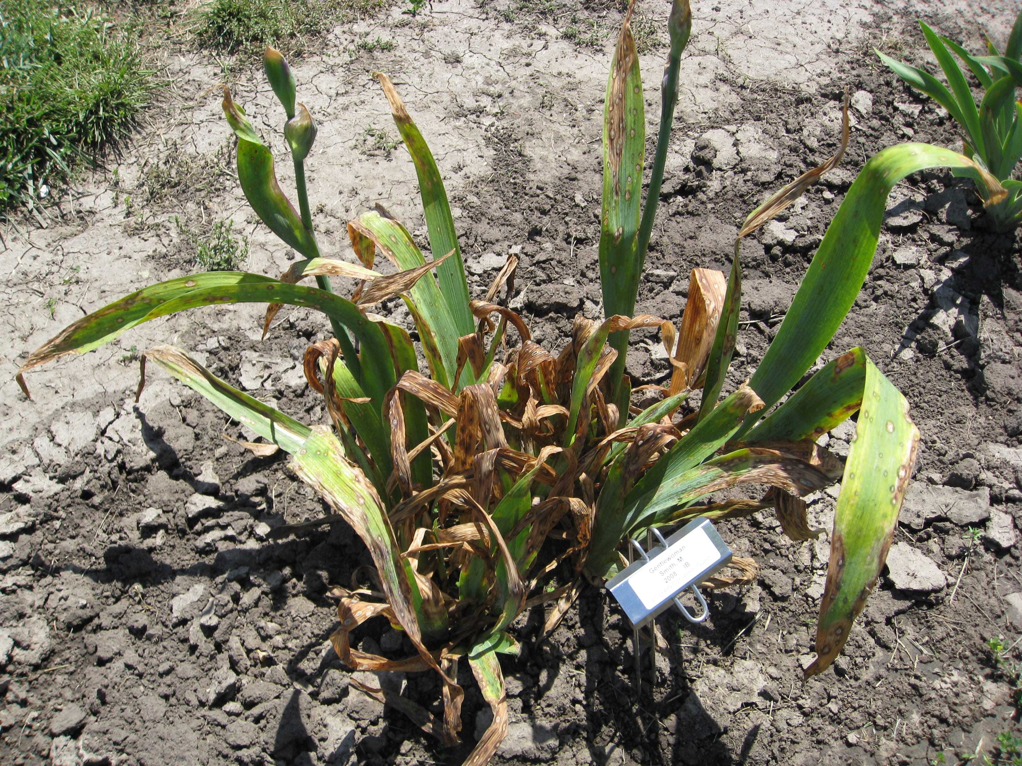
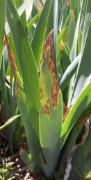
Iris leaf spot is not an easy disease to clean up because it overwinters in the residue. So the first step for management is to clean up the flower bed in the fall after frost has killed the tops. This will help to reduce the amount of disease that is carried over. Unfortunately it won’t get rid of the disease. If the planting is old and crowded, digging them up and respacing them will improve air flow. This can help to reduce disease severity.

Start fungicide protection (chlorothalonil or mycloblutanil) when leaf spotting first shows up early in the spring. Four to six applications may be needed at 7-10 day intervals. Adding a spreader sticker will help coverage and effectiveness of the treatment.

For more information on iris leaf spot check out the following K-State Horticulture fact sheet. https://hnr.k-state.edu/extension/info-center/common-pest-problems/common-pest-problem-new/Iris%20Leaf%20Spot.pdf

Natural needle drop, or plant disease?

(Megan Kennelly, KSU Plant Pathology)

Which of these is a disease, and which is natural needle drop?

If you guessed natural needle drop for the first and disease (Dothistroma needle blight) for the second, you are right!

If you were not sure, here are some resources to figure it out.

In a recent article in Horticulture News, Ward Upham mentioned some recent reports about natural needle drop on evergreens. You can read more about it here:

http://www.ksuhortnewsletter.org/newsletters/natural-needle-drop-on-spruce-arborvitae-and-pines

In addition, this publication talks about pine diseases and at the end there is a section about natural needle drop:

https://www.bookstore.ksre.ksu.edu/pubs/l722.pdf

If you see yellowing, browning, or dropping of needles and you still are not sure you can reach out to your local K-State Research and Extension office or contact the KSU Plant Disease Diagnostic Lab:

Plant Disease Diagnostic Lab

1712 Claflin Road
4032 Throckmorton PSC
Manhattan, Kansas 66506
(785) 532-5810
Fax: (785) 532 5692
clinic@ksu.edu


Large Patch Evaluation Study Update

By: Manoj Chhetri

With temperatures cooling down and days being shorter, we are already starting to see warm-season grasses, including zoysiagrass, going to sleep. At the Rocky Ford Turfgrass Research Center, located in Manhattan, KS, we have shut down the irrigation in warm-season plots.

Our zoysiagrass in the large-patch tolerant breeding plot is not cooperating with us as much as we wanted. We inoculated the field in mid-September with fresh Rhizoctonia pathogen and kept the field pretty wet to encourage fungi to flourish. However, to our dismay, we did not see much of the disease activity, except in a few poor drainage spots. With disease research, it is the type of research where we want disease pathogens to have no mercy on us. We are impatiently waiting for spring, which in fact is a more favorable time of the year for large patch activity.

We are hopeful that we have at least one or two new zoysiagrass progeny that possess greater large patch tolerance. Again, it is hard to make comparison and evaluate when we don’t have disease pressure. So far, we have narrowed down to 10 best progeny out of 60. On the positive note, we have seen more disease pressure on our non-selected progeny than in our top-ten selected progeny. This tells us that we did a good job on choosing those ten-best progeny.

This project is aiming to develop a large patch tolerant zoysiagrass that can significantly reduce cost on fungicides and protect the environment. It is a collaborative project between Texas A & M and K-State University.

Pictured Above: Zoysiagrass progeny evaluated in large patch disease environment.

Pictured Above: One of the zoysiagrass progeny showing large patch in one inoculated half (right side) and fungicide treated cleaner side on other half (left side).

Temperature fluctuations and turf diseases

(Megan Kennelly, KSU Plant Pathology)

As I write this on Thursday we are looking into a weekend with forecast lows deep down into the 30’s. This comes after a September with multiple days in the 90’s and even more in the upper 80’s. Depending on how the weather shakes out after this weekend our warm-season turf may start showing signs of dormancy. Right now it is all still quite green.

In our cool season turf I’ve not seen much dollar spot lately, and it should be simmering down. I have heard a few reports of rust,

and my first suggestion in those cases is to review the fertility regime and make sure it’s not too low on N.

We are seeing and hearing reports of large patch across the region. Here is one example from today at our research facility in Manhattan:

Spring dead spot happens in … spring. But fall is a time to think about it. I’ll point you to an excellent summary of fall disease info from Dr. Miller next door in Missouri, with details about large patch, spring dead spot and other diseases:

https://turfpath.missouri.edu/reports/2019/10_09_19/


Zoysia breeding line evaluation work continues

(Megan Kennelly, KSU Plant Pathology)

KSU continues its work with the turf breeding team at Texas A&M and colleagues at Purdue. We recently inoculated some breeding lines with the large patch pathogen. We grow the fungus on sterilized oats then bury it just under the thatch layer. Sometimes we see symptoms in fall, but often we do not see them until spring. Scientific research takes a lot of patience :). In the meantime, we are keeping the plots moist to foster fungal growth.

In the meantime, large patch is active especially in wet areas:

Take note of these areas. It’s too late to fertilize zoysia now, but when spring comes around a bump of slow-release N may prompt recovery. In the meantime there may be actions you can take to improve drainage.

Breaking disease life cycles with good sanitation

(Megan Kennelly, KSU Plant Pathology)

Many plant pathogens like to survive the winter in infected crop debris. One example is iris leaf spot.


Here is a zoom – the black spots are structures where the fungus produces spores:

Here are some other flower diseases that carry over on debris:

Rose black spot lesions, close-up of lesions, and microscopic view showing abundant spores:

Cercospora leaf spot on hosta:

For many of these common flower-garden leaf spots, removing diseases and dead leaves can help reduce the amount of the fungi lurking over winter to cause disease next year. If composting, don’t put the compost back in the spot where you took the leaves.

Keep an eye out for powdery mildew in your landscape…

By Christian Webb

As summer starts winding down, you may begin to notice some of your bedding plants developing a white powdery coat on the upper surface of their leaves. No, this is not your plants putting on a winter coat for the cool autumn nights. It’s actually caused by the infection of a fungus on the surface of your plants leaves called powdery mildew.

Powdery mildew can be commonly found developing on many types of landscape plants during the late summer. On most plants, this disease will develop on the lower leaves and move up through the canopy. The typically symptoms of the infection will appear as a white to grey dusty growth on the upper surface of the leaves. In severe cases, leave may appear distorted and the cause defoliation.

Pictured to the left: Powdery mildew on Ox-eye Daisy (Heliopsis helianthoides)

To develop, the fungus requires high relative humidity and temperatures around 70⁰F. Many woody and herbaceous ornamentals may be affected by powdery mildew. As a general rule, the powdery mildew on one host will only affect hosts in that genus. For example, the fungi that you will find on oaks trees will only affect oaks and the powdery mildew on grapes will only affect grapes.

If you are looking to control powdery mildew in your landscape, there are a few ways to protect your plants. For annuals, increasing the spacing between plants will increase airflow through the canopy create more unfavorable conditions for powdery mildew develop. Along with altering the plant canopy, consider planting resistant hosts. Replanting the same susceptible hosts will likely result in a reinfection.

Pictured above on left: Powdery mildew on Zinnia (Zinnia elegans). Right: A microscopic image of the powdery mildew spores on the surface of a Zinnia leaf.

There are labeled fungicides for the control of powdery mildew. But you consider apply any product, be sure that your infection is caused by powdery mildew. Downy mildews looks similar to powdery mildew, but are caused by a different type of organism and may not be controlled by products labeled for powdery mildew. Powdery mildew is typically found on the upper surface of the leaf, while downy mildew is associated with the underside of the leaf. For recommended products for the control of powdery mildew, please consult the following factsheet: Link.

For further assistant in identifying powdery mildew, contact your local extension office.

To find your local extension office, the list of Kansas extension offices can be found here.

Further discussion of powdery mildew on woody ornamentals can be found at this Link on pgs. 48-50

Reference:

https://www.fs.fed.us/rm/pubs/rmrs_gtr335.pdf

https://hnr.k-state.edu/extension/info-center/common-pest-problems/common-pest-problem-new/Peony%20Powdery%20Mildew.pdf

Powdery Mildew Resistant Peony Varieties

By Kenneth Dodson

Powdery Mildew is a common fungal disease that can appear on a wide range of plants. This year has seen a high level of powdery mildew on peonies, leaving bushes looking snow-covered or dusty. The best way to avoid dusty looking peony bushes is to use resistant plants. A look through the peony demonstration planting at the K-State Gardens showed a wide range of disease susceptibility to powdery mildew.

Pictured above: Powdery mildew on Peony (Paeonia spp.) 

Here are a few peony varieties that showed good resistance to powdery mildew. In terms of Herbaceous Peonies: Red Charm, Lillian Wild, and Pink Luau were the best options, followed by Madame de Verneville, and Pink Hawaiian Coral. If you’re looking for Tree Peony varieties, there are several that show strong resistance to powdery mildew: Rockii, Joseph Rock, Kamata Nishiki, Shima Daijin, Hakuo Jishi, Kinkaku, and Kokamon all looked good. Lastly, the Itoh or intersectional peonies Pea Green, Yellow Charm, and Morning Lilac exhibited good disease control against powdery mildew.

Just fyi, the major distinction between the three growing types is the in the way they overwinter. Tree peonies will have a woody stem that stays above ground, while herbaceous peony stems will die back to

the ground every year. Itoh varieties have herbaceous stems as well, and will die above ground, but Itoh peonies typically have the same flowering types as tree peonies.

Ash Leaf Spot on Green Ash Trees in Kansas

By: Christian Webb

You may notice brown spots beginning to develop on the lower foliage of green ash trees. Symptoms of Ash leaf spot include irregularly shaped brown spots on the leaf that merge to form large patches of brown spots. In severe cases, the tree may defoliate four to six weeks earlier than normal causing the tree to appear bare long before other trees lose their leaves.

Pictured above on the left: A branch of lower leaves on an Ash tree affected by leaf spot. Right: Brown leaf spots with white speckling on the surface of the upper leaf caused by Ash leaf spot

The fungi that cause Ash leaf spot (Mycosphaerella spp.) survive in the leaf litter underneath the ash tree. In the early summer, the fungi in the leaf litter will begin producing spores capable of infecting leaves. Initial infections will appear on the lower leaves, and then quickly spread throughout the tree. By late fall, the fungus will produce structures able to survive the harsh conditions of winter and be ready to infect new leaves during the following summer.

The severity of the disease will depend on the conditions present during the period of leaf infection. Typically, the disease will become apparent in the late summer and early fall, which may not cause significant cosmetic damage. But in summers with abnormally wet conditions, the disease may develop earlier than normal and cause premature defoliation. Multiple years of these conditions may lead to weak trees with reduced vigor prone to branch dieback.

Pictured above: A microscopic image of fungal structure emerging from the surface of the leaf.

There are few recommended methods to protect ash plantings from Ash leaf spot. Since the disease will survive in the residue around the trees, raking up fallen leaves will reduce the amount of disease carried over to the next growing season. No ash trees resistant to leaf spot have been reported although they may vary in susceptibility.

Reference:

More information on Ash leaf spot may be found at the following Link on page 46-47. https://www.fs.fed.us/rm/pubs/rmrs_gtr335.pdf